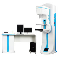
WMC-RC-X8500C HF Digital Radiography System
This machine is applied to take radiography on every part of human body, such as head, limbs, chest, limbus and abdomen and etc.
Features
- Type of generator and X-ray tube:
- Flat Panel Detector
- Apply with A-Si (Amorphous silicon) Toshiba imported Flat panel detector, which could give perfect digital images directly.
- 3K×3K Acquisition matrix, 143um pixels size, and 3.6Lp/mm ultimate spatial resolution, with DQE values≥ 66%
- Shortest imaging time ≤ 15s
- 17〞×17〞Large acquisition area and with the non-center processing technology, no matter the center and border, the quality of the image is the same.
- The detector could be rotated ±45 degree along the axis direction, to satisfy the different photograph requirement of every body parts, such as Ankle joint, lateral spine
- The detector has the self-protection function. It can stop to move when it detect the distance in front of the barrier.
- Digital Working Station.
- Operation System.
- Be equipped with 19〞imported LCD high resolution monitor screen,the delicate and richness degree of image is far higher than the normal medical monitor. International advanced level.
- Brightness and Contrast are higher than 1000NIT, far higher than the normal LCD screen of 400 NIT.
- These features can make the doctor diagnose more accurate and smooth.
- Human graphical touchable screen. Just need a slight click on the human body position and shape, the parameters can be set easily.
- Be equipped with the microphone and remote exposure control. The doctor can control outside the operating room.
- Be equipped with various set of infrared facilities to protect the machine from the mis-operation of the doctors.
- Optional PLXF153 operating room. Battery power supplied, Infrared unlock
- Optional SONY, CODONICS film printer.
- Mechanical Movement:
- The self-designed and manufactured electric U-arm mainframe can move up and down, and rotate in a wide range, which can satisfy the requirements of multi-site photography.
- Adopting original Italian geared motor, the features are reliable performance, lower noise, longer service life.
- The unique three three-dimension and three independent control system, can achieve one-key reposition.
Specification
| High-frequency X-ray machine | Output power | 50kW | |
| Main inverter frequency | 60kHz | ||
| X-ray tube | Dual-focus X-ray tube | Small focus:0.6 Large focus:1.2 | |
| Output power | 27kW/75 kW | ||
| Anode Capacity | 210kJ(300kU) | ||
| Anode Angle | 12° | ||
| Speed of rotating anode | 9700rpm | ||
| Tube Current | 10mA- 500mA | ||
| Tube voltage | 40-150kV | ||
| mAs | 0.1-630mAs | ||
| Exposure Time | 0.001-12.5s | ||
| AEC | Yes | ||
| Digital Image System | Digital Detector | Field of view | 17”*17” |
| Pixel | 3K*3K | ||
| Ultimate spatial resolution | 3.6LP/mm | ||
| Pixel size | 143um | ||
| Output grayscale | 14bit | ||
| Imaging time | 15s | ||
| Image Workstation | Acquisition module | Inside enhancement module | |
| Image information management | Dicom image transmission | ||
| Dicom film printing | |||
| Dicom image storage (hard disk, compact disk) | |||
| Mechanical structure and performance | U-arm | Vertical movement range | ≥1250 mm(motorized control) |
| Focus-screen movement range | 1000mm-1800mm(motorized control) | ||
| Rotation range | -40°-+130°(motorized control) | ||
| Detector rotation | -40°-+40° | ||
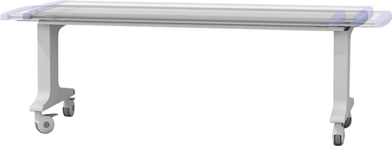
| Photography table (Optional) | Table size | 2000mm*650mm | |
| Table height | ≤740mm | ||
| Transverse movement | 200mm(electromagnetic lock) | ||
| Longitudinal movement | 100mm(electromagnetic lock) | ||
| Power supply | 380V 50/60Hz | ||
Options
PLXF153Intelligent All-Directions Mobile Table, electromagnetic control of floating table surface, chargeable.
(2000×650×740mm, Longitudinal movement: 200mm, Horizontal movement:100mm)