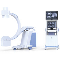
WMC-RC-D5600B High Frequency Digital X-Ray Radiography with Portable Flat Panel Detector
And meet different positions radiography, such as lay decubitus, normotopia, side position etc.
1. Original imported high frequency host and high speed tube
- TOSHIBA highest inverter frequency and high voltage generator
Constant direct current high voltage output, it can achieve high quality monochromatic X-Ray and eliminate the harmful effect of soft ray to image thoroughly. - Equipped with high performance and large capacity X-Ray tube
- Adopt 0.6/1.2mm2 dual focus, 300KHU large capacity and high speed X-Ray tube,it is suitable for long time and high intensity clinical examination.
- Intelligent high voltage control system with multiple human APR, LCD display show APR rules, and set according patients figure, position and part automatically, it’s fast and convenient.
- Computer progress control, remote operate, electric multi-lead collimator, adjust X-ray range as diagnosis need, this reduce X-ray harm for patients and atmosphere
- Have IBS, there are four choices to set KV and mA diagram. AEC offer accurate exposure for different figures and promise lowers radiation for high definition photos.
- Multi self-diagnosis program, has RS232 port, and with error indication function.
2. High definition digital image chain to ensure that minor lesions can not be passed
- Imported DR flat panel detector, it does not need any conversion but it can finish digital photography of each position to achieve real direct digital.
- DR flat panel detector DR connected with image processor achieves low noise and rich contrast image, edge enhancement filtering device makes the edge of image more clear and sharp.
- 3k×3k gathering matrix, more than 900 mega pixel, promise high photo space definition, and good signal-no-noise ratio.
- 14”*17” portable, and 17”*17” fixed flat panel detector can cover whole abdomen and whole chest, no limitation for photography.
- Fast image speed, work together with DROC soft, and promote work efficiency.
- A-si stable function, low requirement for temperature, and no crystal under high temperature.
3. Equipped with new digital image system achieves rich digital image processing function
- Advanced digital image processing function
- Pre-assembled Windows XP professional operating system and professional image processing software
- Image playback: thumbnails view, sequence replay tools.
- Image processing: W/L adjustment, arrows and words etc labels, angle and distance measurement, image scaling, translation, up/down conversion, left/right conversion, rotation, black and white reversal.
- Image storage: image real-time storage, DICOM image send, A shampoo Burning Studio, derived storage (choose various storage ways Bitmap, JPEG, AVI etc to be used in Word and Power-point office software, it is convenient for the doctors to write diagnostic reports and papers).
- DICOM3.0: can be connected with laser camera to print film and PACS NETWORK.
- Medical record management: database management, graphic report, support for WORKLIST.
4. Comfortable, convenient and exquisite table
- Larger SID range can meet whole body such as head, chest, abdomen, waist, limbs and different positions radiography, such as lay decubitus, normotopia, side position etc.
- Two bed options, SYC20 and SYC30, SYC20 is normal bed ,SYC30 is elevator bed ,can move up and down, give extra long range, this promise wheelchair and stretcher radiography. And more up and down range, this way is more convenient for small figure.
5. Bucky stand
- Standard normal bucky stand , can upgrade to electrical bucky stand , this kind bucky stand can move automatically with X-ray tube ,don’t need doctor to operate the bucky stand manually, make operation much easier and save more time.
Main Technical Parameters
| tem | Content | Technical Parameters | |||
| Power | Voltage | 380 V±38 V | |||
| Frequency | 50 Hz±1 Hz | ||||
| Capacitance | ≥65 kVA | ||||
| Internal resistance | ≤0.17Ω | ||||
| High Frequency High Voltage X-Ray Generator | Power | 50 KW | |||
| Inverter frequency | 30 KHz | ||||
| Tube voltage | 40 kV~ 150 kV, continuous adjustment | ||||
| Tube current | 10 mA~ 630 mA, stepping adjustment | ||||
| Exposure time | 1.0 ms~ 6300 ms, stepping adjustment | ||||
| mAs range | 0.2 mAs~500 mAs | ||||
| X-ray source assembly | Focal spot | 1.2 mm /0.6 mm | |||
| Input power | 50 kW/20 kW | ||||
| Anode thermal capacity | 380 KHU | ||||
| Rotary anode speed | 2800 rpm | ||||
| Bucky stand | Up/down range | ≥1300 mm | |||
| Grid | Density: 130 L/inch | ||||
| Ratio: 10:1 | |||||
| Focal distance: 180 cm | |||||
| Size: 15” x 18” | |||||
| Stand column | Tube/CCD center to floor distance | 1150 mm | |||
| Platform horizontal movement | ±120 mm | ||||
| X-ray tube SID | 1000 mm | ||||
| Rotatable foot paddle | ±360° | ||||
| Collimator | Electric multi leaf | ||||
| Flat panel detector | TOSHIBA portable | Material: CsI | |||
| Resolution: ≥3.7l p/mm | |||||
| Valid area: 350 mm(H)×430 mm(V) | |||||
| Pixel matrix: 2448 (H)×2984 (V) | |||||
| Pixel pitch: 143 μm | |||||
| Toshiba Digital Flat Penal Detector | Toshiba | FDX3543RP | |||
| Active area | 350(H)×430(V) | ||||
| Pixel matrix: | 2448(H)×2984(V) | ||||
| Pixel pitch: | 143 μm | ||||
| Cycle time: | 6S | ||||
| Limiting resolution | Min. 3.7 line pair/㎜ | ||||
| A / D transition: | 16 bit | ||||
| Energy range | 40 – 150 kVp | ||||
| Maximum entrance dose (low gain) | 4 mR / frame | ||||
| Data output | 16-bit digital output Ethernet(1000BASE – T) | ||||
| Exposure Synchronous control: | outside | ||||
| power Input: | DC 24V 2A | ||||
| Bed | Bed size | 2000*760mm | |||
| Bed longitudinal movement stroke | ≥900mm | ||||
| Bed longitudinal movement stroke | ≥220mm | ||||
| Bed height from the ground | ≤700mm | ||||
| Bed to film distance | ≤60mm | ||||
| Longitudinal travel of the radiator | ≥500mm | ||||
| Box loading specifications | 8”*10”~14”*17” | ||||
| Fixed filter grid | South Korea microgroove, Focusing distance: 130cm, Grid density: 103L/INCH, Electric vibrator | ||||
| Longitudinal travel of the ball | ≥1300mm | ||||
| Tube column rotation range | +180°·-180° | ||||
| The focus of the tube to the film SID | 500~1280mm | ||||
| The tube assembly rotates around the arm | ≥±90° | ||||
| Speed governor | Manual multi-leaf style | ||||
| The beam light is turned on for a limited time | 15s | ||||
| Image acquisition workstation | Color LED display | Resolution: 1280 x 1024 | |||
| Size: 19” 1M | |||||
| Brightness: 250 cd/m2, | |||||
| Contrast: 1000:1 | |||||
| Ratio: 5:4 | |||||
| Pixel pitch: 0.294 mm | |||||
| Gray to gray response time: 5 ms | |||||
| Viewing angle: 160°/170° | |||||
| DR0C workstation software | Basic operation: change user password, edit user, acquire image | ||||
| Additional operation: add new checks, edit current checking information, add new positions, change image acquiring sequence, switch between multiple checking protocols, manual adjustment of exposure parameters, AEC, select focus, select patient’s body type, check tube heat content, select ESA curve, clip image, add remark (the remark will be sent to DICOM workstation), add position mark to image, rotate or flip image, view image in full size, check information of patient and exposure dose, accept or refuse image. | |||||
| Image management: change list sequence order, edit patient’s basic information after exposure, check history image, resend history image, reprint history image, check additional image information, preview history image, manage refused image, space recycle strategy, protect image, manually delete image | |||||
| System management: edit user, change user password, emergency registration set, check statistics information, detector calibration, equipment control, output queue management, image measurement. | |||||